
Paper Read-throughs, Episode 26 · Takahashi & Yamanaka 2006 · Cell 126(4):663-676 · DOI 10.1016/j.cell.2006.07.024
A question I had before reading the paper
Have you ever wondered why a wound heals into a scar, not brand-new skin?
A skin cell gets damaged, and the body can only patch it up. It cannot regenerate the original tissue from scratch. Once a stem cell differentiates into a skin cell, it stays a skin cell forever. Cell differentiation is a one-way street – you can drive forward but never U-turn.
That was my understanding of cell fate before I read this paper.
In 2006, a team at Kyoto University published a paper in Cell. Fourteen pages. It proved one thing:cell fate is not a one-way street. Four genetic switches are all it takes to send an ordinary skin cell back to its embryonic state.
This year marks the paper’s twentieth anniversary. Six years after publication, it won the Nobel Prize.
Today we walk through this paper: what they did, how they did it, and what it means for you.
Why the old path did not work
Before 2006, scientists already knew about a cell that could become anything in the body – the embryonic stem cell. These cells had two remarkable abilities: they could multiply indefinitely while retaining the potential to turn into any cell type.
The problem: harvesting them required destroying embryos. This raised ethical issues and limited supply. If a patient needed their own cells for therapy, there was also the rejection problem – someone else’s cells trigger an immune attack.
Could we make these cells from a patient’s own ordinary tissue?
That question had haunted the field for years. Existing evidence pointed to two paths: transferring an adult cell’s nucleus into an egg (cloning), or fusing an adult cell with an embryonic stem cell. Both proved that some kind of “reset mechanism” exists inside cells. But nobody knew which factors drove it.
This paper’s entry point was right there.
Not addition, but subtraction
The researchers’ logic was straightforward.
Since certain genes are especially active in embryonic stem cells and maintain their “master cell” properties, what if you put those genes into ordinary cells? Could you send them back?
They started by picking 24 candidate genes. These 24 were either highly expressed in embryonic stem cells or had been linked to stem cell properties by earlier research.
Then they did something I find elegant:not addition, but subtraction.
First, they loaded all 24 genes into ordinary mouse embryonic fibroblasts (structural cells from skin tissue, essentially). Could these cells become stem-like? Yes. Twenty-two colonies survived, and some looked exactly like embryonic stem cells – round, large nucleoli, scant cytoplasm.
Next, they removed genes one at a time and watched which removal killed the colony.
Like sending 24 team members home one by one. Whose departure breaks the project? That person is the key.
The beauty of this experiment is that it does not ask “what do we need to add?” but “who can we not do without?”
After rounds of elimination, the answer emerged:only four factors were needed – Oct3/4, Sox2, Klf4, and c-Myc.
With just three, cells survived but looked nothing like stem cells and could not become anything else. One short and the whole thing falls apart. You only need to hear those names once; from now on I call them “the four switches.”
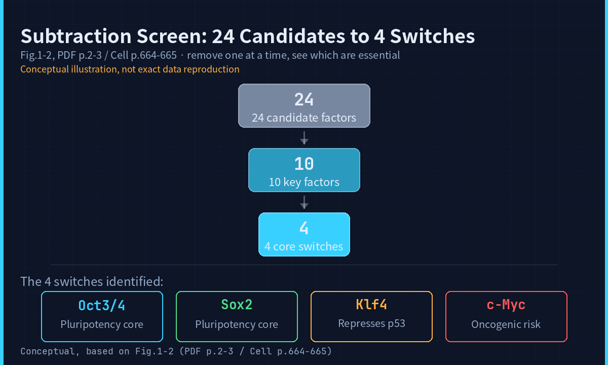
My reading: these four were not random picks. Oct3/4 and Sox2 were already known as core transcription factors maintaining the “pluripotent” state in embryonic stem cells. The surprise was the two “helpers” – Klf4 and c-Myc, both genes associated with tumors. An oncogene participating in cell reprogramming was unexpected at the time.
Hard evidence: the reset cells really can become anything
Looking the part is not enough. How do you prove these reset cells have truly regained the ability to become any cell type?
The researchers did something very direct: they injected the cells under the skin of mice.
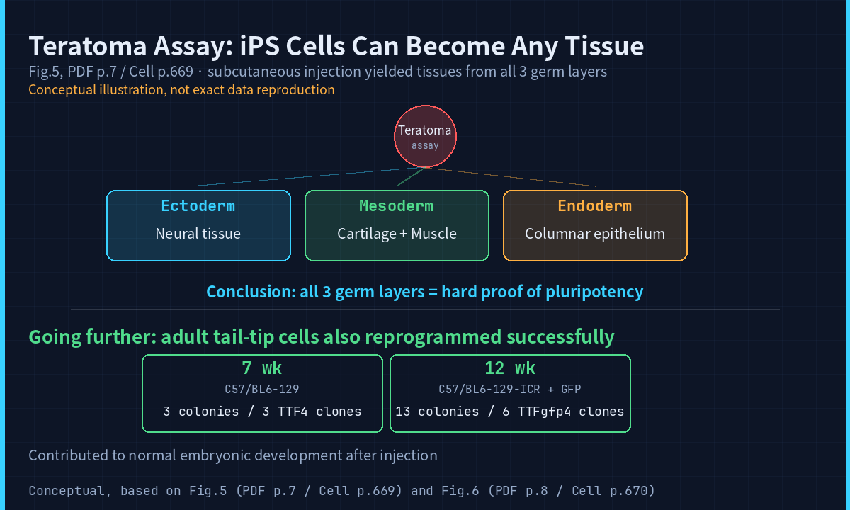
A special tumor grew – a teratoma.
Picture this: a lump of tissue containing nerves, cartilage, and muscle. These completely different tissue types, all from a tube of skin cells. Cut it open, and all three germ layers were represented: ectoderm (neural tissue), mesoderm (cartilage and muscle), endoderm (columnar epithelium).
The tumor was not the goal. It was the proof test. It demonstrated that the reset cells had truly regained the ability to become anything.
More interestingly, they did not stop at embryonic cells.
The team took skin cells from the tail tips of adult mice – not embryos, not pups, but fully grown animals aged seven and twelve weeks. Added the same four switches. The adult cells reverted too. Injected into mouse embryos, they joined normal development and differentiated into tissues of all germ layers.
Even the cells of a grown animal can be sent back.That is one of the most striking findings in this paper.
The paper also reports a chimera experiment: out of 22 embryos at E7.5, 3 showed GFP-positive iPS cell contribution. Out of 18 embryos at E13.5, 2 confirmed iPS cell participation. The numbers look modest, but the significance is huge – it proves the reset cells do not just survive in a dish. They can participate in real development.
However, of 27 pups born, none were confirmed as chimeric mice. The paper’s explanation is candid: silencing of Oct3/4 in the iPS cells was not complete, which may explain the failure to obtain live chimeras. This kind of honest reporting was uncommon at the time.
The honest caveats: similar, but not identical
This next section is what I respect most about this paper.
The researchers did not claim the reset cells were identical to real embryonic stem cells. They performed whole-genome expression profiling (DNA microarray analysis). The result:
The two were similar, but not identical.
Specifically, some genes were turned on at lower levels in the reset cells. The paper classified genes into three groups: Group I, highly expressed in both ES and iPS cells (Myb, Kit, Gdf3, Zic3); Group II, highly expressed in ES cells and high-quality iPS but not in low-quality iPS (including Nanog, Sox2); Group III, highly expressed only in ES cells (including Dnmt3a, Dnmt3b).
This distinction matters. It tells us that reprogramming is not a simple “factory reset” but an incomplete, layered process. Some switches were successfully reset; others were not.
There was also the viral vector issue. The retroviruses used to deliver the four switches left about 20 copies in each cell’s genome (the Southern blot analysis showed roughly 20 integration sites per clone). The cell’s DNA was colonized by foreign gene fragments. Fine for research, a concern for clinical use.
Another easily overlooked detail: these iPS cells could not remain undifferentiated without feeder cells, even with LIF. This meant they depended more heavily on culture conditions than embryonic stem cells did. The paper listed this as another difference.
These details define the boundaries of the technology – where the path is already cleared, and where the hazards remain.
The cancer risk: a hurdle that cannot be ignored
One of the four switches is called c-Myc. It is an oncogene. Its normal job is to push cells to grow and divide. When it is too active, the result is cancer.
The paper states clearly: “Use of c-Myc may not be suitable for clinical applications.”
That warning was not theoretical. The paper itself reported that some mice injected with iPS cells developed tumors. In the teratoma experiment, tumors from iPS-MEF3 clones were composed entirely of undifferentiated cells with no signs of differentiation at all.
The paper’s analysis of the cancer mechanism is worth reading. The c-Myc protein associates with histone acetyltransferase (HAT) complexes, including TRRAP, CBP, and p300. In the mammalian genome, c-Myc may bind to as many as 25,000 sites – far more than Oct3/4 or Sox2. c-Myc might induce global histone acetylation, allowing Oct3/4 and Sox2 to access their specific targets. That is a clever mechanistic hypothesis, but it also means c-Myc’s reach is too broad – it does not just flip the switches you need. It starts fires everywhere.
The paper also describes an interesting balance: Klf4 directly represses p53 (a tumor suppressor), while c-Myc suppresses p21 (a cell cycle inhibitor). During reprogramming, the two may form a mutual check – but if that balance breaks in the body, the consequence could be tumor formation.
Later teams replaced c-Myc or used safer delivery methods (such as Sendai virus or plasmids instead of retroviruses). Those improvements were made by others, not by this paper – do not credit them to this work.
That is the full picture of this paper: a breakthrough and a risk. Both written in the same fourteen pages.
From paper to Nobel Prize: what six years means
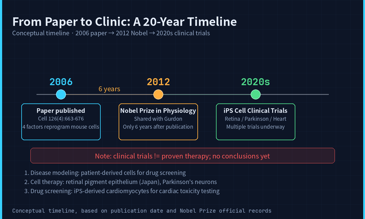
This paper was published in 2006. The Nobel Prize in Physiology or Medicine came in 2012.
Six years.
In academia, that speed means everyone immediately understood how important this was. Nobel Prizes usually wait twenty or thirty years for an impact to be fully validated. Awarding it in six says the committee thought the significance needed no further waiting.
Today, scientists use these cells to build disease models, screen drugs, and develop therapies. Japan has launched clinical trials using iPS-derived cells for eye diseases. Multiple other trials are underway worldwide.
Note my phrasing: underway. I am not claiming they cure anything. Those studies are still in progress, with no conclusions to report.
The “stem cell shot” question
You may have seen ads for “stem cell anti-aging shots.” Some cost tens of thousands of yuan per injection, promising to make people younger, to reverse aging.
Here is what you need to know.
The science is solid. This paper is one piece of evidence, and multiple clinical trials are real. The field has moved fast since 2006. New trials keep launching.
But some products sell research-stage technology as proven therapy. Those are different things. Promising in research does not equal proven effective.
I cannot give medical advice about any brand or treatment. What you can do is distinguish between solid science and packaged marketing.
A few points worth flagging
After reading this paper, there are several things I want to highlight.
First, reprogramming efficiency is extremely low.The paper itself acknowledges that only a small fraction of cells expressing all four factors became iPS cells. They offered several explanations: the four factors need to land in a precise expression range (the paper cites data showing that a mere 50% increase or decrease in Oct3/4 protein induces differentiation of ES cells); additional chromosomal alterations may be required; or retroviral integration in certain clones may have happened to activate the right gene combination. Efficiency remains a core challenge in this field today.
Second, the origin of iPS cells is still debated.The paper discusses the possibility that iPS cells might not come from direct reprogramming of fibroblasts but from rare tissue stem cells already present in the culture (about 0.067% of skin cells are stem cells). But they argued against it: inducing iPS from bone marrow stroma (which should contain more mesenchymal stem cells) was equally inefficient, and cells induced by only three factors were completely nullipotent. These observations do not support the “tissue stem cell origin” hypothesis, though they do not settle the question either.
Third, the four factors interact in subtle, mutual-restraining ways.c-Myc promotes proliferation; Klf4 represses p53 but activates p21 (a cell cycle inhibitor); c-Myc suppresses p21. The paper states: “the balance between c-Myc and Klf4 may be important for the generation of iPS cells.” These four switches do not work independently – they have complex interactions. This insight informed later improvements to reprogramming protocols.
Fourth, the paper’s final paragraphs mention a direction:they found that a Nanog reporter system could replace Fbx15 for selection. This meant they already had ideas for improving the screening method – using stricter criteria might yield higher-quality iPS cells. Subsequent research took that direction much further.
Three things to remember
Remember the question we started with? Why does a wound heal into a scar instead of regenerating perfect skin?
Because cell differentiation is a one-way street – or so we thought before 2006.
This fourteen-page paper proved:four switches can reset an ordinary skin cell to factory settings.That is not a metaphor. It is experimental fact.
First, cell fate is not a one-way street. Four genetic switches can send a mature cell back to its embryonic state.
Second, this is not science fiction. Clinical trials using iPS-derived cells are underway. Not yet proven therapies, but the direction is clear.
Third, not all stem cells are equal. There is a clear line between what the science shows and what some ads claim.
We are the kind of people who read papers all the way to the end. If you want to know how these four switches reached the hospital, subscribe. We will see you next time.
Reference
Takahashi, K. & Yamanaka, S. (2006). Induction of Pluripotent Stem Cells from Mouse Embryonic and Adult Fibroblast Cultures by Defined Factors. Cell, 126(4), 663-676. DOI: 10.1016/j.cell.2006.07.024